Small Animal Models
Animal research plays a vital role in understanding health and disease, which is essential for developing medicines and therapies for people and animals. Animals are only used in research at Glasgow when there is no other suitable alternative available, and we are committed to the principles of the three Rs – replacement, refinement, and reduction.
To date, we have conducted Syrian hamster and mouse models to study SARS-CoV-2 pathogenesis and immune responses, and to examine in vivo efficacy of antiviral compounds, assessed infection of IFNAR KO mice with Severe fever with thrombocytopaenia syndrome virus (SFTSV, Dabie bandavirus) and Bhanja virus (BHAV) evaluating any clinical signs and immune responses/markters, harvest tissues and blood for veterinary pathology studies.
Our Syrian hamster models provide an excellent platform for the assessment of SARS-CoV-2 infection. As hamsters are naturally susceptible to SARS-CoV-2 infection, they do not require humanisation. This allows for the assessment of virulence in the context of the whole organism rather than just the isolated cell and organoid models. Mice are not naturally susceptible to SARS-CoV-2 infection; however, we are able to provide studies using humanised mice as required. In these animals, human ACE2 expression is driven either through adenoviral transduction in the respiratory tract or through genetic modification.
We intend to expand our capabilities, subject to legal, regulatory and Home Office approval, to support the research efforts around addressing potential future viral threats including developing models for influenza virus and adenovirus/adeno-associated virus.
Further information on how animals are used in research, our policies, legislation and guidance are available on the University of Glasgow - Research website.
Modes of Infection
Primary Infection: Animals infected intranasally with SARS-CoV-2
Reinfection: Animals reinfected intranasally with the same or different SARS-CoV-2 variant 3 weeks after primary infection
Transmission: Animals infected passively with SARS-CoV-2 through direct contact with infected animals or through air exchange
Assessment of Disease
Throat, nose or rectal swabs can be collected daily to assess viraemia
Weights, temperatures and disease scores recorded daily
Pathology
Custom solutions for every experimental pathology project
Histopathology diagnosis of animal tissues and organs by a veterinary pathologist (Diplomate of the European College of Veterinary Pathologists)
Quantification of morphological findings
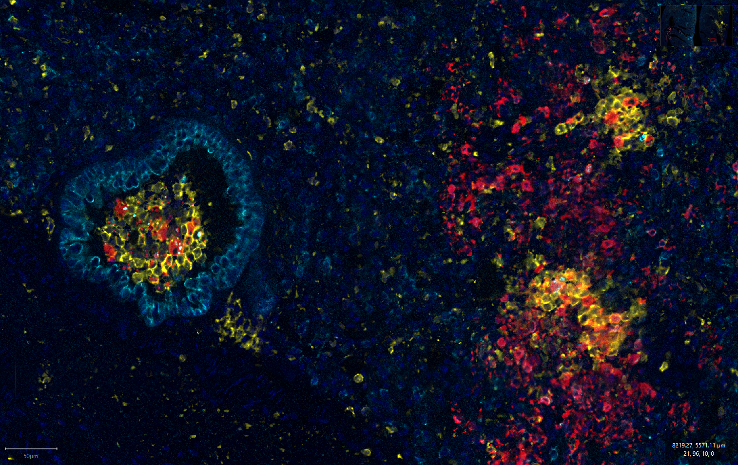
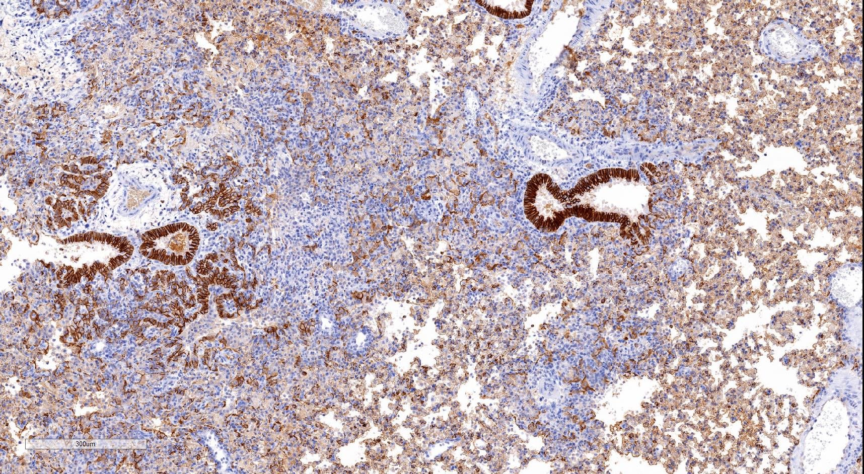
Immunohistochemistry and immunofluorescence for immunophenotyping and detection of viral or cellular proteins of interest
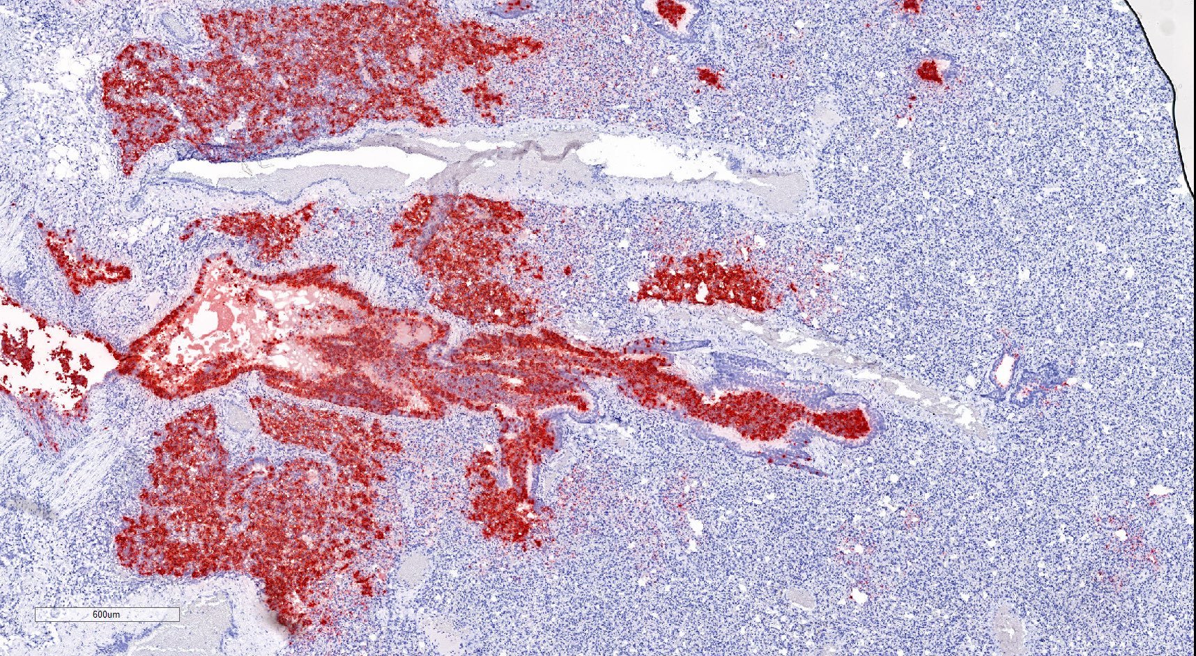
In situ-hybridisation (bright field and fluorescent; including multiplex staining) to detect viral and cellular RNA interest
Software-assisted quantification (HALO, Aperio, QuPath) of immunohistochemistry, immunofluorescence and in situ-hybridisation of whole scanned slides
Scanning and archiving of whole microscopic slides
Deep phenotyping of tissue microenvironment (PhenoCycler)
Spatial transcriptomics using the Nanostring GeoMx-technology
Tissue Processing
Techniques can be tailored to the needs of any study
Cell based assay to measure live virus (plaque assays, foci forming unit assays)
Multiplex RT-qPCR for measuring viral RNA in different tissues
RNA Sequencing (bulk or single cells)
Measure of tissue and serum cytokines
Live cell sorting for downstream analysis (limited to mouse studies)
Flow cytometry to measure immune response (limited to mouse studies)
Analysis of serum antibodies
Statistical analysis provided of all results
Compounds Screening
Able to screen drugs, antibodies, vaccines and immunomodulators
Delivered via intranasal, IP, IV, SC, or oral (gavage or in food/water) routes
Compounds administered up to twice daily
Option of osmotic minipump implantation for continuous compound delivery


Additional Services
Breeding, maintenance, and housing of specialised transgenic animals within our facility as required
Viral loads can be measured throughout the study through the use of throat swabs and nasal washes with other measurements including weight, temperature and clinical scores, recorded daily.
We employ a number of techniques for tissue processing including RT-qPCR, RNA sequencing, histology, cytokine and other cell based assays, cell sorting and flow cytometry and antibody analysis. These techniques can all be tailored to the needs of your study.
